
Transesophageal Echocardiogram (TEE): Procedure, Benefits, Risks & Recovery
A transesophageal echocardiogram, often called a TEE, is a heart ultrasound performed from inside the esophagus rather than from outside the chest. This position gives doctors a much closer, clearer view of the heart’s structures.
Because the esophagus sits directly behind the heart, sound waves travel a much shorter distance than they would through skin, muscle, and ribs. This gives sharper images, especially for structures like heart valves and small blood clots that a standard chest ultrasound can sometimes miss.
This guide covers why doctors order a TEE, how to prepare, what happens during the procedure, and what your results mean.
What Is a Transesophageal Echocardiogram (TEE)?
A transesophageal echocardiogram uses a thin, flexible probe with an ultrasound transducer on the tip. The probe passes through the mouth and down the esophagus, positioning it directly behind the heart.
Standard ultrasound imaging works by sending sound waves into the body and reading the waves that bounce back to build a picture. During a TEE, this happens from just behind the heart rather than through the chest wall, which normally blocks some of the image quality due to ribs, lung tissue, and body size.
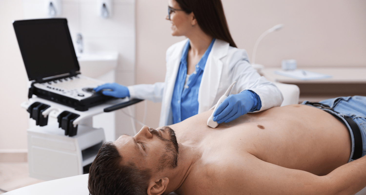
This differs from a standard echocardiogram, also called a transthoracic echocardiogram, which uses a probe moved across the outside of the chest. A TEE gives a closer, more detailed view, particularly useful for structures near the back of the heart.
Why Is a TEE Performed?
Doctors order a TEE for a wide range of reasons.
Heart valve disease. A TEE gives a detailed look at valve structure and function, often clearer than a standard echocardiogram.
Blood clots. The test is especially useful for finding clots in the left atrial appendage, a small pouch in the heart where clots often form.
Stroke evaluation. When the cause of a stroke is unclear, a TEE helps doctors look for a heart based source, such as a hidden clot.
Atrial fibrillation. Patients with atrial fibrillation often need a TEE before certain treatments to rule out clots in the heart.
Infective endocarditis. The test helps confirm or rule out an infection on the heart valves, a condition known as endocarditis.
Congenital heart disease. A TEE can give detailed views of structural heart defects present from birth.
Aortic disease. The test helps evaluate the aorta for problems such as an aortic dissection or aortic aneurysm.
Cardiac tumors. A TEE can help identify masses within the heart chambers.
Prosthetic valve assessment. For patients with an artificial heart valve, a TEE gives a clearer picture than external imaging alone.
Before cardioversion. Doctors often order a TEE before cardioversion to confirm no clots are present before restoring a normal heart rhythm.
During structural heart procedures. A TEE can guide doctors in real time during certain heart procedures.
Monitoring during heart surgery. Surgeons sometimes use a TEE during surgery to watch heart function continuously.
Who Needs a TEE?
A TEE is often recommended for patients with:
- Suspected heart valve problems
- A stroke with an unclear cause
- Suspected blood clots in the heart
- Signs of a heart infection
- Congenital heart abnormalities
- Poor quality images from a standard transthoracic echocardiogram
TEE vs Transthoracic Echocardiogram (TTE)
| Feature | TEE | TTE |
| Image quality | Very high, especially for valves and clots | Good, but can be limited by body size or lung tissue |
| Invasiveness | Semi invasive, probe passes through the esophagus | Non invasive, probe stays on the chest |
| Sedation | Usually required | Not required |
| Preparation | Fasting required | No fasting required |
| Best uses | Detailed valve and clot evaluation | General heart structure and function screening |
| Risks | Low, but slightly higher than TTE | Very low |
| Accuracy | Very high for specific conditions | High for general screening |
| Recovery | Short monitoring period after sedation | None needed |
Doctors often start with a standard TTE and move to a TEE only when a closer look is needed, such as when clot detection or detailed valve assessment is required.
How to Prepare for a TEE
Preparing properly helps the procedure go smoothly and safely.
- Fasting is usually required for about 6 hours before the test.
- Follow your doctor’s instructions about regular medications.
- Diabetes medication may need adjustment, based on your physician’s guidance.
- If you take a blood thinner, your doctor will give specific instructions beforehand.
- Remove dentures and jewelry before the procedure.
- Arrange for someone to drive you home afterward, since sedation is used.
- Tell your provider about any esophageal disease, swallowing difficulties, allergies, pregnancy, or previous reactions to sedation.
What Happens During the Procedure?
Check-in. You arrive and complete registration before the test.
Consent. Your care team reviews the procedure and answers any questions before you sign consent.
IV placement. A small IV line is placed to deliver sedation medication.
Monitoring. Sensors track your heart rate, blood pressure, and oxygen levels throughout the test.
Local throat anesthetic. A numbing spray is applied to your throat to ease probe insertion.
Sedation. Medication through the IV helps you relax and stay comfortable during the test.
Probe insertion. The doctor gently guides the probe through your mouth and down your esophagus.
Ultrasound imaging. The probe captures detailed images and Doppler blood flow data from behind the heart.
Probe removal. Once imaging is complete, the probe is carefully removed.
Recovery. You rest under observation while the sedation wears off.
The full procedure usually takes between 30 and 60 minutes, including a short recovery period for monitoring afterward.
Will the Procedure Hurt?
Most patients feel only mild discomfort. A gagging sensation is common as the probe passes down the throat, though sedation and the throat numbing spray help minimize this. Some people notice a temporary sore throat afterward, which usually resolves within a day.
What Does a TEE Detect?
A TEE gives detailed information about several heart structures and conditions:
- Heart valve conditions, including mitral valve disease, aortic valve disease, and tricuspid valve disease
- Blood clots, especially those forming in the left atrial appendage
- Infective endocarditis affecting the heart valves
- Septal defects between heart chambers
- Aortic aneurysm or dissection
- Cardiac masses or tumors
- Complications involving prosthetic heart valves
- Congenital heart defects
Benefits of TEE
A TEE offers several clear advantages over other imaging options:
- Excellent image quality, especially for structures near the back of the heart
- Better detail than a standard echocardiogram for certain conditions
- Real time imaging that supports quick clinical decisions
- Ability to guide certain heart procedures directly
- High diagnostic accuracy for valve disease and blood clots
- No radiation exposure involved
Risks and Possible Complications
Complications from a TEE are uncommon, but patients should be aware of the possibilities, including:
- Sore throat
- Minor bleeding
- Rare esophageal injury
- Reactions to sedation
- Aspiration
- Irregular heartbeat during the procedure
- Rare dental injury during probe placement
Contact your doctor right away if you notice severe throat pain, difficulty swallowing, chest pain, fever, or signs of an allergic reaction after the test.
Recovery After a TEE
After the procedure, you will stay under observation until the sedation wears off. Most people can eat and drink again once their throat numbness fades and their swallowing reflex returns to normal, usually within a few hours.
You should not drive for 24 hours after sedation. Most people return to normal daily activities the next day, though your doctor will give specific guidance based on your individual case.
Understanding TEE Results
Normal findings show healthy valve function, no blood clots, and no structural abnormalities.
Abnormal findings may include:
- Valve disease affecting how well blood moves through the heart
- Vegetations, which are clumps of infection material on a valve
- Blood clots, particularly in the left atrial appendage
- Septal defects between heart chambers
- Reduced heart pumping function
- Aortic abnormalities such as an aneurysm or dissection
Depending on the findings, your doctor may recommend further testing, medication, or a procedure to address the issue directly.
Limitations of TEE
A TEE does not replace every type of heart imaging. It requires sedation, which carries its own small risks. Certain esophageal conditions may make the procedure unsafe, so your doctor will screen for these beforehand. Results also depend somewhat on operator experience, since probe positioning affects image quality. In some cases, doctors may also use imaging such as a cardiac MRI scanner or cardiac CT scanner alongside a TEE for a fuller picture.
Final Thoughts
A transesophageal echocardiogram gives doctors one of the clearest possible views of the heart’s valves and chambers, helping catch problems that other imaging tests can miss. This makes it a valuable tool for evaluating unexplained strokes, valve disease, and heart rhythm problems such as arrhythmias.
Detailed cardiac imaging like this fits into a much larger picture of long term heart health. The BaleDoneen Method combines advanced imaging with a full look at root causes and risk factors, supporting a complete approach to stroke prevention and long term cardiovascular care.