
Carotid Ultrasound: Purpose, Procedure, Results & Benefits
A carotid ultrasound checks the blood vessels in your neck that carry blood to your brain. It uses sound waves, not radiation, to create a picture of these arteries and the blood moving through them.
Doctors order this test to look for narrowing or plaque buildup that could raise the risk of a stroke. Since strokes often happen without warning, this simple scan gives doctors a way to spot trouble early, before a bigger problem strikes.
This guide covers what the test shows, why doctors recommend it, how to prepare, what happens during the scan, and how to understand your results.
What Is a Carotid Ultrasound?
A carotid ultrasound is an imaging test that uses sound waves to check the carotid arteries in your neck. A technician moves a small device called a transducer across your skin, and the machine turns the returning sound waves into a picture of your artery walls and the blood flowing through them.
The test also measures how fast blood moves through these arteries. This helps doctors spot narrowing, called stenosis, or areas where plaque has started to build up along the artery wall.
What Are the Carotid Arteries?
The carotid arteries run along both sides of your neck. You can often feel their pulse just below your jaw. These arteries carry oxygen rich blood from your heart up to your brain.
Because the brain depends entirely on this blood supply, any blockage or narrowing in these arteries can seriously affect brain function. A significant blockage can reduce blood flow enough to cause a stroke, which is why doctors pay close attention to the health of these vessels.
Why Is a Carotid Ultrasound Performed?
Doctors order a carotid ultrasound for several reasons tied to stroke prevention.
Checking for artery disease. The test looks for signs of carotid artery disease, where plaque narrows the artery over time and reduces blood flow to the brain.
Finding plaque buildup. Plaque linked to atherosclerosis can build up slowly along the artery wall, and this test shows how much has accumulated.
Measuring narrowing. The scan measures how much the artery has narrowed, known as stenosis, which helps guide treatment decisions.
Checking for blood clots. Clots can form near plaque and break loose, traveling to the brain and causing a stroke.
Evaluating reduced blood flow. The test shows whether blood is moving through the artery at a normal speed or if flow has slowed due to a blockage.
Following up after a TIA. After a transient ischemic attack, doctors often order this scan to check whether narrowed arteries played a role.
Monitoring known disease. Patients already diagnosed with carotid artery narrowing may repeat the scan over time to track any changes.
Who Needs a Carotid Ultrasound?
Doctors often recommend this test for people with:
- High blood pressure, including diagnosed hypertension
- High cholesterol
- Diabetes
- A history of smoking
- Existing heart disease
- A previous stroke
- A family history of stroke or artery disease
- An abnormal sound heard over the carotid artery during a physical exam, known as a carotid bruit
Symptoms That May Lead to Testing
Certain symptoms often prompt a doctor to order a carotid ultrasound, including:
- Temporary vision loss in one eye
- Sudden dizziness
- Sudden weakness on one side of the body
- Facial drooping
- Trouble speaking
- Numbness or tingling
- A mini stroke, also called a TIA
If you notice any of these symptoms, review the full list of warning signs and symptoms and contact your doctor right away.
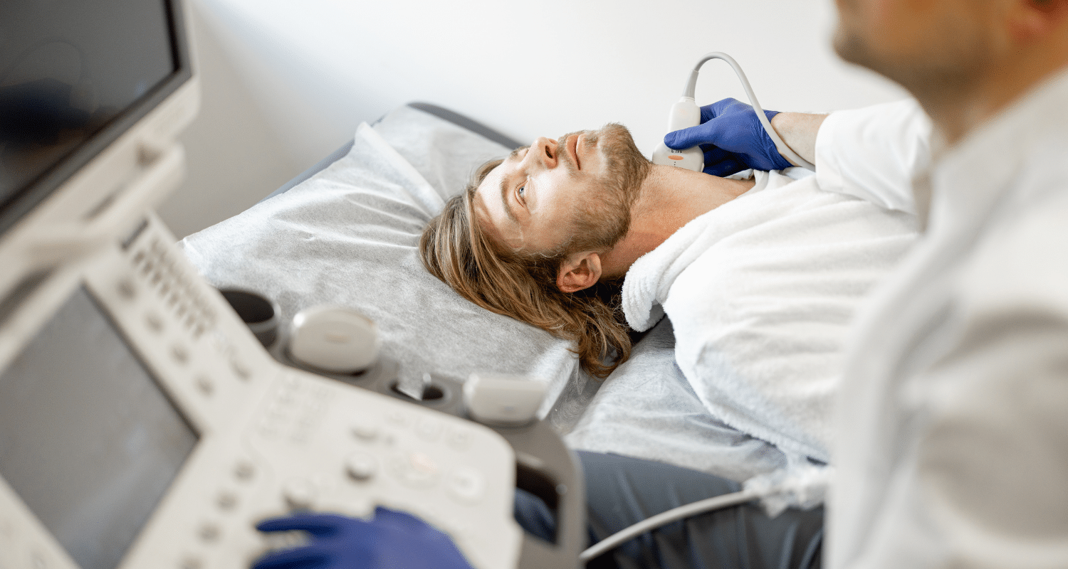
How Does a Carotid Ultrasound Work?
The test uses high frequency sound waves sent into your neck through a handheld transducer. These waves bounce off the artery walls and the blood cells moving through them, then return to the machine, which turns this data into a live image.
A version called Doppler imaging measures the speed and direction of blood flow. When combined with the standard image, this is known as a duplex ultrasound, since it captures both the structure of the artery and how blood moves through it. This same Doppler approach is used in other parts of the body too, including tests like vascular ultrasound used to check circulation elsewhere.
The images also reveal plaque along the artery wall, giving doctors a direct look at buildup that might otherwise go unnoticed.
Types of Carotid Ultrasound
Standard ultrasound creates a basic image of the artery structure.
Doppler ultrasound adds information about blood flow speed and direction.
Duplex ultrasound combines standard imaging with Doppler data for a complete view of both structure and flow.
Color Doppler imaging adds color to the image, making it easier to see the direction and speed of blood flow at a glance.
How to Prepare
Preparing for a carotid ultrasound is simple.
- No fasting is required.
- Wear comfortable clothing that allows easy access to your neck.
- Remove necklaces or other jewelry around your neck before the test.
- Continue taking your regular medications unless your doctor tells you otherwise.
What Happens During the Procedure?
Registration. You check in and confirm your information before the test begins.
Gel application. A technician applies a small amount of gel to your neck, which helps the sound waves transmit clearly.
Probe movement. The technician gently moves the transducer along both sides of your neck to capture images from different angles.
Image capture. The machine records still images and video of your artery structure.
Doppler measurements. The technician also records blood flow speed at several points along the artery.
Duration. The full test usually takes between 30 and 45 minutes.
Is the Test Painful?
No. A carotid ultrasound is completely painless. It is non invasive, meaning nothing enters your body, and it uses no needles and no radiation at all. Most people feel nothing more than light pressure from the transducer against their skin.
Understanding Carotid Ultrasound Results
Normal results show healthy blood flow with no significant narrowing in either carotid artery.
Abnormal results may show:
- Mild plaque buildup with little effect on blood flow
- Mild stenosis, a small amount of narrowing
- Moderate stenosis, where blood flow is noticeably reduced
- Severe stenosis, where the artery has narrowed significantly
- Complete blockage, where no blood flow passes through that section of the artery
Your doctor reviews the percentage of narrowing along with your symptoms and other risk factors before deciding on next steps.
What Happens If Blockages Are Found?
Treatment depends on how severe the narrowing is and your overall health.
Lifestyle changes often come first, including diet changes, exercise, and quitting smoking.
Blood pressure control helps reduce further strain on the artery walls.
Statins, often discussed under statins and cardiovascular risk, help manage cholesterol and slow further plaque buildup.
Blood thinners, sometimes part of antiplatelet therapy, help reduce the risk of clot formation.
Surgery may be needed for severe narrowing. A carotid endarterectomy removes plaque directly from the artery, while carotid artery stenting places a small mesh tube to hold the artery open.
Benefits of Carotid Ultrasound
This test offers several clear advantages:
- Early detection of artery narrowing before symptoms appear
- Strong support for stroke prevention efforts
- A safe procedure with no known risks
- No radiation exposure
- Easily repeatable over time to track changes
- Wide availability at most imaging centers and hospitals
- Fast results that support quick decision making
Limitations
The test does have some limits. Results can depend on the skill of the technician performing the scan, since positioning and technique affect image quality. Some patients, particularly those with a shorter or larger neck, may be harder to visualize clearly. In some cases, doctors may order a CTA or MRA to confirm findings. The test also cannot diagnose every possible vascular condition on its own.
Are There Any Risks?
A carotid ultrasound carries no meaningful risk. There is no radiation, no injections, and no recovery time needed afterward. This makes it one of the safest imaging tests available for checking artery health.
Carotid Ultrasound vs Other Imaging Tests
| Test | Radiation | Invasiveness | Best For |
| Carotid ultrasound (duplex) | None | Non invasive | First line screening |
| CT angiography (CTA) | Yes | Requires contrast dye | Detailed anatomy confirmation |
| MR angiography (MRA) | None | May require contrast | Detailed imaging without radiation |
| Cerebral angiography | Yes | Invasive, catheter based | Precise mapping before surgery |
| CIMT | None | Non invasive | Measuring early artery wall thickness |
Carotid ultrasound remains the usual first step because it is safe, quick, and widely available. When more detail is needed, doctors may add a CTA, MRA, or a test like CIMT for a fuller picture of early arterial changes.
How Much Does a Carotid Ultrasound Cost?
Cost varies depending on where you have the test done. Hospitals often charge more than standalone imaging centers. Most insurance plans, including Medicare, cover carotid ultrasound when a doctor orders it for a medical reason. Out of pocket costs also vary by region, so it helps to check with your insurance provider and the testing center beforehand.
Final Thoughts
A carotid ultrasound gives doctors a safe, clear look at the arteries that supply blood to your brain. Finding narrowing or plaque early gives you and your doctor time to act before a stroke happens.
This test works best as part of a complete plan for stroke and heart health, one that also looks at cholesterol, blood pressure, and other risk markers together. The BaleDoneen Method combines tools like carotid ultrasound with advanced cardiovascular testing to build a full picture of your risk and support long term stroke prevention.